
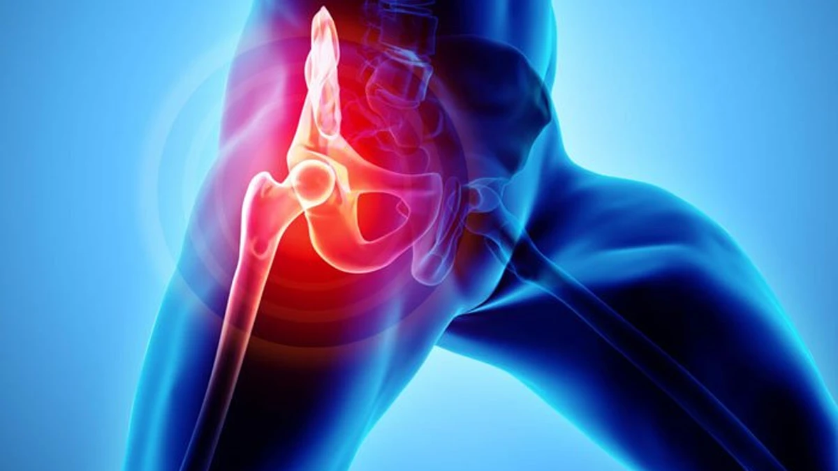
What is avascular necrosis?
Contrary to what is supposed, bone is not a lifeless structure; it is living tissue nourished by blood vessels. When the blood flow to a region is cut off or reduced, the bone cells in that region die. This is called avascular necrosis.
The most frequently affected region is the head of the thigh bone within the hip joint. Because its blood supply comes from a limited number of vessels, it is more open to disturbances of nourishment.
Dead bone tissue keeps its shape for a time. For this reason there may be no complaint in the early period. In time the bone surface becomes unable to carry the load placed on it and collapses; after this stage the joint cartilage is also rapidly damaged.
Why does it develop?
In a proportion of cases no cause can be established. The known risk factors include the following:
- Trauma: Hip dislocation and fractures of the neck of the thigh bone can injure the vessels supplying the region.
- Long-term corticosteroid use: The risk increases with high-dose and long-term use.
- Alcohol use: Regular and heavy use is counted among the risk factors.
- Certain blood disorders: Conditions such as sickle cell anaemia can affect the blood supply to bone.
- Decompression sickness: Vessel blockage related to changes in pressure.
Carrying a risk factor does not mean the condition will develop; equally, it can also be seen in people with no risk factors.
Symptoms and diagnosis
At an early stage there is most often no symptom. The findings that appear as it progresses:
- Pain in the groin that increases over time, at times radiating to the outer side of the hip and the inner side of the knee
- Pain that becomes pronounced on weight-bearing and walking and eases with rest
- Night pain at an advanced stage
- Restriction of hip movement and limping
An X-ray may appear normal at an early stage. Where there is suspicion, an MRI is requested; MRI can show the disturbance of blood supply in the bone tissue before any change appears on X-ray. For this reason a normal X-ray is not on its own sufficient when clinical suspicion exists.
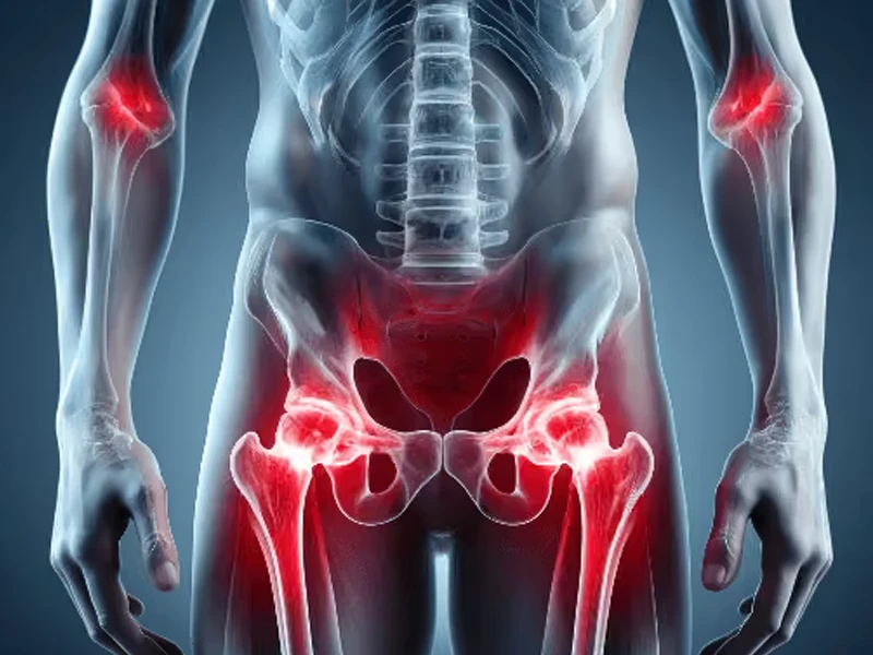
Treatment options
Treatment is determined according to the stage of the condition and whether the bone surface has collapsed.
At an early stage (while the surface has not yet collapsed) the aim is to preserve the joint:
- Reducing load and modifying activity
- Reviewing any risk factor that is present
- Joint-preserving procedures directed at reducing pressure within the bone and supporting the blood supply
At an advanced stage (once the surface has collapsed and the joint is damaged) the chance of joint-preserving methods diminishes; at this stage hip replacement comes onto the agenda. For other problems of the hip region you can see the hip surgery page.
Examination and follow-up process
-
History and physical examination
The duration and nature of the pain, the risk factors and the range of hip movement are assessed.
-
Imaging
It begins with X-ray; if suspicion persists, early-stage changes are investigated with MRI.
-
Staging and plan
The state of the bone surface is established; a joint-preserving approach or the replacement option is discussed.
-
Follow-up
At an early stage, progression is monitored with regular imaging and the plan updated accordingly.
Frequently asked questions
Does avascular necrosis resolve on its own?
My X-ray came back normal — does that mean I do not have avascular necrosis?
I take cortisone — will I develop avascular necrosis?
Is avascular necrosis seen at a young age?
Can it occur in both hips at once?
Is a replacement always necessary in avascular necrosis?

Treating physician
Prof. Dr. Melih Malkoç
Specialist in Orthopaedics and Traumatology. Graduate of Ege University Faculty of Medicine; completed his specialist training at Şişli Etfal Training and Research Hospital and received the title of associate professor in 2015.
Full biography →The information on this page is for general guidance only and does not replace a diagnosis or treatment recommendation. Diagnosis and treatment decisions are made by the physician on an individual basis following examination and imaging.